A Reconditioning Hub Deep Dive
Everyone Is Looking at Connor Bedard's Shoulders. I'm Looking Further Back.
Connor Bedard has experienced three shoulder injuries in less than a year, including two remarkably similar right shoulder incidents during faceoffs.
In December 2025, he injured his right shoulder during a faceoff against St. Louis. The injury was managed without surgery.
In July 2026, he sustained a separate left shoulder injury after falling awkwardly into the boards during offseason training. That injury required surgical intervention.
Then, on November 17, 2026, another faceoff against Toronto resulted in a further right shoulder injury.
The mechanisms are interesting.
Both right shoulder injuries occurred during faceoffs, when unexpected contact with an opponent's stick placed a sudden demand on his shoulder. The left shoulder injury involved a more conventional traumatic mechanism, a fall into the boards.
Naturally, attention has focused on the shoulders themselves. Structural instability, labral integrity, surgical management and rehabilitation are all important considerations.
But I am interested in an injury that occurred nearly three years ago.
In January 2024, Bedard sustained a fractured mandible following a hit against the New Jersey Devils. The injury required surgical fixation.
From my perspective, that injury deserves considerably more attention when examining his subsequent shoulder problems.
I believe significant mandibular trauma has the potential to produce lasting changes in the neuromuscular relationships between the jaw, cervical spine, thoracic cage and shoulder girdle.
Those changes may affect how the shoulder is positioned, stabilized and prepared to respond to external forces.
My clinical hypothesis is that residual dysfunction following Bedard's mandibular injury could be contributing to his shoulder vulnerability, particularly on the right side.
His recurrent right shoulder instability clearly involves structural considerations. But structural damage and altered neuromuscular function are not mutually exclusive.
In fact, understanding how they interact may be essential to developing a more complete reconditioning strategy.
And while his left shoulder injury involved a separate traumatic mechanism, I would also want to understand whether any broader functional adaptations were present before that injury occurred.
The question is not simply why Bedard's shoulder became unstable. It is whether his entire shoulder complex was functioning optimally before the injury occurred.
That requires looking beyond the glenohumeral joint.
Why Bedard's Injury History Raises Questions
The shoulder is often discussed as though it operates independently.
We examine the labrum, capsule, ligaments, rotator cuff and surrounding musculature. We identify structural pathology and develop a rehabilitation plan intended to restore stability and strength.
All of this is important.
But the glenohumeral joint is only one component of a much larger functional system.
Its stability depends on the coordinated interaction of the scapula, clavicle, thoracic cage, cervical spine and neuromuscular system.
The scapula must provide an appropriate foundation for the humerus. The thoracic cage must allow the scapula to move effectively. The cervical region contributes to the muscular and neurological systems involved in controlling this relationship.
And the jaw is functionally connected to the cervical region.
Following a significant mandibular fracture, I would be interested in several potential adaptations.
Did the injury alter mandibular mobility or resting jaw muscle activity?
Was there a change in chewing patterns, clenching behavior or temporomandibular function?
Did the trauma or subsequent surgery produce persistent sensory changes, tissue restrictions or altered cervical muscle recruitment?
Could any of those adaptations influence scapular control or the timing of muscular stabilization during high-speed movement?
These questions become particularly relevant when considering Bedard's right shoulder injuries.
A faceoff involves a rapid interaction between the upper extremities, trunk and lower body. The athlete must generate force while responding to unpredictable external resistance.
The shoulder must be capable of both producing movement and maintaining joint stability.
If the muscles responsible for positioning and stabilizing the shoulder are not coordinating effectively, the joint may be less prepared to accommodate sudden loading. One detail from Bedard's original injury particularly caught my attention.
Following the hit, Bedard reportedly described his face as feeling numb and his bite being off.
Those are two potentially important clinical findings.
Facial numbness raises questions about trigeminal sensory involvement, while an altered bite suggests a disruption in mandibular alignment or occlusal function following the trauma.
From a reconditioning perspective, I would want to know whether these symptoms completely resolved and, more importantly, whether normal sensory and motor function was restored.
Altered sensory input can lead to altered motor output.
If significant mandibular trauma disrupted sensory information from the jaw and surrounding tissues, could that have contributed to persistent adaptations in cervical muscle recruitment and shoulder-girdle control?
This is precisely why I believe Bedard's previous jaw injury deserves closer examination.
Was Healing the Primary Focus of His Jaw Rehabilitation?
Another aspect of Bedard's recovery interests me.
Fracture rehabilitation, particularly following mandibular fixation, often places considerable emphasis on protecting the healing bone, managing pain, restoring essential jaw function and progressing activity as healing permits.
Those priorities are appropriate.
But I wonder how comprehensively the functional consequences of his original trauma were assessed.
Was his recovery primarily guided by fracture healing and symptom resolution, or did it also involve a detailed assessment of the temporomandibular joints, cervical spine, cranial structures and associated myofascial relationships?
Were there residual restrictions involving the mandible or surrounding tissues?
Was cervical motor control examined following the injury?
Did anyone assess whether changes in jaw function influenced shoulder-girdle recruitment?
I am also interested in the intraosseous consequences of the original fracture.
From an osteopathic perspective, trauma involves more than the visible fracture line. It also affects the periosteum, surrounding soft tissues and the local mechanical environment of the bone.
Surgical fixation introduces another consideration.
A plate creates a localized region of increased structural stiffness, and depending on its position, it may influence the mechanical behavior of the repaired area.
In my clinical interpretation, retained fixation hardware may also complicate the assessment or treatment of perceived intraosseous restrictions. This is an osteopathic observation rather than an established explanation for distant musculoskeletal dysfunction.
I would want to know whether fixation hardware remains in place and whether any residual restrictions or symptoms are associated with the repaired region.
The bone may be structurally healed, but has the entire functional system recovered?
That is the distinction I believe deserves greater attention.
Other Professional Athletes Have Raised Similar Questions
Bedard is not the first elite athlete whose craniofacial or dental history has prompted questions about musculoskeletal function.
Two professional football cases are particularly interesting.
Kevin De Bruyne: Facial Trauma and Recurrent Muscular Injuries
During the 2021 Champions League final, Kevin De Bruyne sustained fractures involving his nose and left orbital region following a collision with Chelsea defender Antonio Rüdiger.
He subsequently experienced significant hamstring problems, including injuries during the 2023 Champions League final and the opening match of the following Premier League season. He underwent hamstring surgery in August 2023.
In April 2025, former professional footballer Joey Barton publicly suggested that dental treatment De Bruyne had received might be contributing to his injury problems.
The theory attracted criticism and was widely treated as unconventional.
I think it deserved a more thoughtful discussion.
Dental procedures, prolonged jaw opening, changes in occlusion and temporomandibular dysfunction can influence aspects of mandibular and cervical function.
That provides a legitimate anatomical and physiological basis for investigating potential relationships beyond the oral cavity.
With De Bruyne, however, I would actually be more interested in his previous nasal and orbital fractures.
Significant trauma to the craniofacial region can affect sensory structures, soft tissues and neuromuscular function.
The trigeminal system has important relationships with cervical sensory processing, and cervical function contributes to broader movement coordination.
I would want to know whether De Bruyne experienced any persistent sensory or cervical changes following his facial injuries.
The connection to hamstring function is less direct than the jaw-cervical-shoulder relationship I am proposing for Bedard.
Nevertheless, I believe the potential influence of altered neuromuscular coordination and posterior-chain function warrants investigation.
The posterior muscular chain is not a single mechanical structure extending from the skull to the hamstrings. It is a functional system involving multiple muscles, joints and neurological relationships.
Changes in head and cervical control can influence trunk movement strategies, while trunk and pelvic mechanics contribute to lower-extremity loading.
Whether a previous facial injury could meaningfully influence that system is an important research question.
The broader point is that the body organizes movement through coordinated systems rather than isolated anatomical regions.
A previous injury may alter that coordination long after the original tissues have healed.
Aly Cissokho: A Debate That Began Years Earlier
In 2009, French footballer Aly Cissokho was preparing to join AC Milan when concerns arose during his medical examination.
Milan officials publicly suggested that abnormalities involving his dental health could contribute to muscular problems and potentially affect his future physical condition.
The proposed transfer ultimately collapsed.
Cissokho disputed the medical explanation and maintained that financial disagreements were responsible.
What interests me is not whether AC Milan was correct about Cissokho.
It is that an elite professional football organization was considering the potential relationship between oral health and muscular function more than 15 years ago.
The subject remains controversial, partly because different mechanisms are frequently grouped together.
Periodontal inflammation, dental occlusion, temporomandibular dysfunction and direct craniofacial trauma are distinct clinical issues.
But they all raise questions about whether the oral and craniofacial systems should be considered more carefully within comprehensive musculoskeletal assessment.
To understand why, we need to examine the anatomy.
The Anatomy: Understanding the Potential Connection
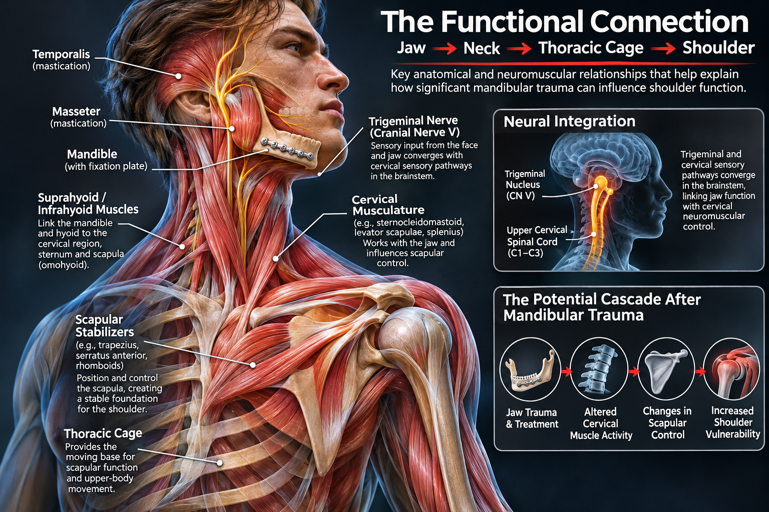
AI-generated anatomical illustration for educational purposes. Anatomical structures and neuromuscular relationships are simplified and may not be precisely represented.
The Trigeminal System and Cervical Spine
The trigeminal nerve (cranial nerve V) is responsible for much of the sensation in the face and jaw. It also controls the muscles involved in chewing and helps the nervous system monitor jaw position and regulate bite force.
What makes this particularly interesting is its relationship with the upper cervical spine.
The jaw and upper neck are neurologically connected. Sensory pathways from these regions overlap within the brainstem and upper spinal cord. This helps explain why problems involving the jaw can sometimes produce symptoms in the head, face and neck, and vice versa.
Following significant mandibular trauma, pain, nerve irritation, altered jaw movement or changes in muscle function may affect the sensory information reaching the nervous system.
And when sensory input changes, motor output may change as well.
From a reconditioning perspective, this raises an important question: Could changes in jaw function following Bedard's fracture have influenced how his cervical muscles coordinate and, potentially, how his shoulder girdle responds to force?
Altered sensory input can lead to altered motor output.
That principle is central to my clinical reasoning.
The nervous system continuously integrates information from muscles, joints, skin and other sensory structures to organize movement.
If significant trauma changes the quality or consistency of that information, the nervous system may adapt its motor strategies.
Those adaptations may initially be protective.
But what happens when the original injury heals and the altered motor strategy persists?
Could the cervical musculature continue to operate differently because of residual changes in mandibular function or sensory input?
And could those changes influence the way the shoulder girdle prepares for and responds to load?
This is one of the principal mechanisms I would investigate in Bedard.
The Mandible, Hyoid and Cervical Musculature
The mandible is controlled by the masseter, temporalis and pterygoid muscles.
These muscles function alongside the suprahyoid and infrahyoid systems, which connect the mandible and hyoid region with the skull, cervical structures, sternum and, through the omohyoid, scapula.
The cervical musculature contributes to head positioning and stabilization during mandibular activity.
Research has demonstrated measurable coactivation between jaw muscles and several cervical muscles during clenching.
These include the sternocleidomastoid, trapezius, levator scapulae and splenius capitis.
This is particularly relevant because the trapezius and levator scapulae also contribute to scapular positioning and control.
The muscles involved in jaw-cervical coordination overlap functionally with muscles responsible for positioning the shoulder girdle.
This creates a potential pathway through which altered mandibular function could influence cervical and scapular motor behavior.
For me, the significance lies not simply in the presence of anatomical connections, but in the way those muscles coordinate their activity.
If mandibular trauma changes the behavior of the masticatory system, what happens to the muscles that participate in both cervical stabilization and shoulder-girdle control?
The Thoracic Cage and Shoulder Complex
The thoracic cage is another important component of this relationship.
The scapula moves over the posterior thoracic wall. Its orientation and movement are influenced by thoracic mobility, rib-cage configuration and the coordinated activity of surrounding musculature.
The trapezius, serratus anterior, rhomboids and levator scapulae all contribute to scapular function.
The shoulder complex must coordinate movement across the sternoclavicular, acromioclavicular, scapulothoracic and glenohumeral articulations.
An athlete may demonstrate adequate strength in isolated testing while still experiencing difficulties coordinating these structures under dynamic loading.
This is why I place considerable importance on examining the entire shoulder complex rather than focusing exclusively on the injured joint.
In Bedard's case, I would be particularly interested in the relationship between cervical function, thoracic mobility, scapular positioning and glenohumeral stability.
A subtle change in the way the scapula is controlled may influence the position and loading demands of the humeral head.
When an unexpected force is applied during a faceoff, the shoulder must respond immediately.
That response depends not only on structural integrity but also on the neuromuscular system's ability to organize and regulate force.
Surgical Trauma, Scarring and Intraosseous Adaptation
Bedard's mandibular injury required surgical fixation.
Depending on the fracture location and surgical approach, mandibular surgery may involve disruption of local soft tissues, periosteum and surrounding muscular or sensory structures.
Healing can involve scar formation, altered tissue mobility and changes in local muscle behavior.
The craniofacial and cervical regions also contain interconnected fascial compartments surrounding muscles, neurovascular structures and visceral structures.
From an osteopathic perspective, these anatomical relationships warrant examination following significant trauma.
I would be interested in the mobility of the mandibular and cervical tissues, the behavior of the masticatory muscles, and whether movement restrictions or sensory changes influence cervical function.
I would also examine the potential intraosseous consequences of the original trauma.
Bone is a living tissue that responds to mechanical loading and undergoes continuous remodeling.
A fracture and subsequent fixation change the local mechanical environment during healing.
Retained metal hardware has different stiffness characteristics from surrounding bone, and this may influence local load distribution.
In osteopathic practice, clinicians may also describe palpable restrictions within or around previously injured bone.
I consider these observations relevant to a comprehensive assessment, while recognizing that the clinical significance of such palpatory findings and their relationship to distant injuries have not been established.
My interest is whether residual changes in the repaired mandibular region could contribute to persistent sensory or muscular adaptations.
Bone healing is one component of recovery. Restoration of coordinated function is another.
What the Research Tells Us
Several areas of research support the physiological relationships underlying this hypothesis.
Facial Trauma Can Have Persistent Functional Consequences
A systematic review by Helmer and colleagues, published online in 2024, examined the relationship between significant direct orofacial trauma and temporomandibular disorders.
The review included 139 studies and identified a consistent association between significant facial trauma and temporomandibular dysfunction in adults.
Importantly, dysfunction could persist long after the initial injury, including following treatment.
This is particularly relevant when considering mandibular fractures requiring surgical fixation.
It demonstrates that successful treatment of the original trauma does not always mean that normal temporomandibular function has been completely restored.
Temporomandibular Dysfunction Is Associated With Cervical Impairment
A 2020 systematic review and meta-analysis examined cervical musculoskeletal function in patients with temporomandibular disorders.
The review included 21 studies.
Patients with temporomandibular dysfunction demonstrated reduced cervical extensor endurance, reduced cervical mobility and greater neck disability compared with controls.
Interestingly, the researchers did not identify consistent differences in craniocervical posture.
I find that distinction important.
The relationship may be less about static posture and more about how the neuromuscular system functions.
For reconditioning, that shifts attention toward mobility, endurance, motor control, sensory integration and the timing of muscle activation.
Jaw Clenching Changes Cervical Muscle Activity
Research by Giannakopoulos and colleagues in 2013 demonstrated neuromuscular interactions between the jaw and cervical musculature during clenching.
Using electromyography, investigators recorded coactivation involving muscles such as the trapezius, levator scapulae, sternocleidomastoid and splenius capitis.
Earlier research by Ehrlich and colleagues also demonstrated increased cervical muscle activity during strong jaw clenching.
These findings are particularly interesting when considered alongside the demands of dynamic shoulder stability.
The jaw can influence muscular activity in regions directly involved in positioning and controlling the shoulder girdle.
Jaw Activity Can Influence Upper-Body Force Production
Research has also demonstrated that jaw clenching can influence muscle activity and force production beyond the jaw itself.
A 2018 study involving 100 healthy young men examined how voluntary teeth clenching and body position affected maximal force production in the upper and lower extremities. Additional research involving competitive roller hockey players investigated the relationship between jaw clenching and upper-body isometric strength.
Together, these studies suggest that jaw activity can influence neuromuscular output during physical tasks.
But producing more force isn't necessarily the same as having better muscular control.
From a reconditioning perspective, I'm more interested in how effectively muscles regulate their activity. Can they activate when needed, coordinate with surrounding muscles and relax when their contribution is no longer required?
If jaw activity can influence muscle recruitment elsewhere in the body, could significant mandibular trauma also influence the timing, coordination or regulation of those muscles?
For an athlete like Bedard, whose shoulder must respond to sudden and unpredictable forces, that distinction could be particularly important.
My Clinical Hypothesis: What Happens When Stabilizing Muscles Cannot Regulate Their Activity Appropriately?
Normal movement depends on the nervous system's ability to recruit muscles with appropriate timing, intensity and coordination.
Muscles must generate force when required, but they must also modulate their activity as movement demands change.
This is particularly important around joints that depend heavily on dynamic stabilization.
The shoulder is one of those joints.
The rotator cuff contributes to humeral head control, while the scapular stabilizers provide a coordinated foundation for upper-extremity movement.
These systems must respond rapidly to changes in direction, speed and external loading.
Now consider the research demonstrating that jaw clenching can increase cervical muscle activity and influence upper-body force production.
What happens if significant mandibular trauma produces persistent changes in jaw muscle activity or clenching behavior?
Could that create an altered resting or task-related recruitment pattern in the cervical and shoulder-girdle musculature?
Could certain muscles become excessively active, or activate at inappropriate times, while others fail to coordinate effectively?
And could that affect the ability of the shoulder complex to respond to unexpected external forces?
My hypothesis is that residual mandibular dysfunction could contribute to altered cervical and scapular muscle recruitment, potentially compromising the timing and coordination required for dynamic shoulder stability.
I am particularly interested in the possibility of excessive or poorly regulated muscle activity.
More muscle activation is not necessarily better.
A muscle that remains excessively active may have difficulty adapting its output to rapidly changing demands. Excessive co-contraction may increase stiffness and alter movement strategies.
The problem may not be that the athlete lacks strength.
It may be that the neuromuscular system is not coordinating that strength efficiently.
This distinction becomes particularly interesting when examining Bedard's two right shoulder injuries.
Both occurred during faceoffs, when his shoulder was exposed to an unexpected force.
If his shoulder girdle was already operating with altered muscular recruitment or reduced adaptability, that could have contributed to the joint's vulnerability.
This is the relationship I believe deserves greater clinical attention.
The same reasoning raises questions about his left shoulder.
Although that injury involved a more conventional traumatic mechanism, altered shoulder-girdle coordination could influence how an athlete responds to a fall or absorbs impact.
The two shoulders do not have to sustain identical injuries for a broader functional impairment to be relevant.
I would want to determine whether Bedard's previous mandibular trauma has left a measurable neuromuscular signature, and whether that signature changes the way his shoulders function.
That is a specific clinical hypothesis that can be investigated rather than dismissed simply because the original injury occurred elsewhere.
Where the Research Is Falling Short
Existing research has established several important relationships between craniofacial trauma, temporomandibular function, cervical neuromuscular activity and muscle recruitment.
But much of this research examines these relationships independently.
We have evidence supporting individual components of the system, but far less research examining how they interact as a whole, particularly following significant trauma and under the demands of elite sport.
Sports-related facial trauma research understandably emphasizes fracture management, surgical outcomes, complications and return-to-play timelines. However, these measures do not necessarily tell us whether normal sensory processing, muscular coordination and functional integration have been restored.
The jaw, cervical spine, thoracic cage and shoulder do not function independently. They share anatomical, neurological and muscular relationships that contribute to coordinated movement and the body's response to external forces.
Yet there is limited research examining mandibular function, cervical muscle recruitment, thoracic mechanics, scapular control and dynamic shoulder stability together, particularly in athletes recovering from significant craniofacial trauma.
For example, research has demonstrated that jaw clenching can influence cervical muscle activity and force production. But what happens to these relationships following a significant mandibular fracture?
Does altered sensory input influence muscle activation timing, coordination or the ability to respond to unexpected forces?
Do these adaptations persist after the fracture has healed and the athlete has returned to competition?
And could they contribute to altered loading or vulnerability elsewhere in the musculoskeletal system?
These are measurable questions, but they require us to examine the integrated system rather than individual structures in isolation.
The Value of Individual Clinical Investigation
Large prospective studies would help us better understand whether craniofacial trauma is associated with subsequent neuromuscular changes or injury patterns. However, meaningful research does not always have to begin with hundreds of athletes.
An individual athlete represents an N=1 clinical investigation.
If an athlete demonstrates persistent mandibular dysfunction alongside altered cervical or scapular recruitment, we can establish baseline measurements, introduce targeted interventions and evaluate whether meaningful functional changes occur.
Does improving mandibular function change cervical muscle recruitment? Does that influence scapular control? Do those changes carry over into dynamic shoulder testing or sport-specific activity?
A carefully documented clinical case cannot establish a universal causal relationship, but it can identify important patterns and generate questions for larger studies.
This is where I believe an important gap exists between our understanding of individual anatomical structures and our understanding of the integrated human system.
We have identified many of the pieces. What we have not adequately investigated is how those pieces influence one another following injury and whether adaptations in one region may contribute to vulnerability elsewhere.
Sometimes advancing our understanding begins with one carefully examined athlete and a question that conventional assessment has not answered.
What I Would Want to Assess in Connor Bedard
If I were involved in Bedard's reconditioning process, I would want to examine his shoulder injuries within the context of his entire injury history.
That would begin with understanding the original mandibular fracture.
Where exactly did the fracture occur? What surgical approach was used? Does fixation hardware remain in place? Were there any postoperative complications? Did he experience changes in sensation, chewing function, jaw mobility or cervical symptoms?
I would then examine several interconnected systems.
Cranial, Mandibular and Temporomandibular Function
I would assess opening and closing mechanics, deviation, muscle activity, symptoms, chewing patterns and potential clenching behavior.
From an osteopathic perspective, I would also examine the cranial base, cranial vault and associated soft-tissue relationships, particularly where previous trauma or symptoms suggest involvement.
I would be interested in the mechanical behavior of the repaired mandibular region, including the possible influence of retained fixation hardware.
These findings would be considered alongside established measures of mandibular function and neurological status.
Cervical Neuromuscular Function
I would examine cervical mobility, muscular endurance, sensorimotor control and recruitment patterns, particularly involving the sternocleidomastoid, deep cervical musculature, trapezius and levator scapulae.
I would also be interested in whether changes in mandibular position or activity influence cervical muscle behavior.
Thoracic and Scapular Mechanics
I would assess thoracic mobility, rib-cage function, scapular positioning and the ability to coordinate scapular movement under progressive loading.
I would be particularly interested in how these structures function together rather than relying exclusively on isolated strength measurements.
Dynamic Shoulder Stability
Structural assessment remains essential, particularly given his recurrent right shoulder instability.
But I would also want to understand how the rotator cuff, scapular stabilizers and trunk respond to rapid movement and unexpected external forces.
Where appropriate, objective testing could examine muscle activation timing, force production, endurance and responses to controlled perturbations.
Sport-Specific Loading
Finally, I would examine the actual demands of hockey.
Faceoffs, shooting, passing, contact and rapid changes in body position place different demands on the shoulder complex.
I would be particularly interested in whether cervical or mandibular positioning influences muscle recruitment or movement quality during these tasks.
The objective would be to identify measurable impairments, determine whether they influence function and develop a strategy to address them alongside the necessary structural rehabilitation.
This is where I believe reconditioning can contribute something important.
It brings the injury history, anatomy, neuromuscular system and sporting demands into the same clinical investigation.
Moving Forward: A Broader Approach to Craniofacial Trauma in Sport
Professional hockey provides a particularly interesting environment in which to study these relationships.
Players are exposed to pucks, sticks, collisions and other mechanisms capable of producing significant facial trauma.
Although protective equipment has improved, the face and jaw remain vulnerable, particularly in players who compete without full facial protection.
Mandibular fractures, dental injuries and other craniofacial trauma are not uncommon within the sport.
Yet the long-term consequences of these injuries are usually discussed in terms of fracture healing, surgical management and return to competition.
I believe we should be asking more questions about what happens afterward.
Are there persistent sensory changes?
Are there changes in mandibular and cervical motor control?
Does the athlete demonstrate altered recruitment of muscles involved in scapular positioning?
And could these adaptations influence the way the athlete tolerates future sporting demands?
These are questions that deserve collaboration between maxillofacial specialists, sports medicine physicians, dentists, athletic therapists, rehabilitation professionals and researchers.
A more integrated approach could help identify whether clinically meaningful relationships exist and, importantly, how to address them.
Before We Debate Surgery, Are We Asking Why the Shoulder Became Vulnerable?
Much of the discussion surrounding Connor Bedard now centers on whether he requires surgery to stabilize his right shoulder.
I understand that discussion. Recurrent instability can involve significant structural damage, and surgical stabilization may ultimately be the appropriate treatment.
But I believe we should also be asking a different question.
Why has his shoulder become vulnerable during a movement he has performed thousands of times throughout his hockey career?
What I find particularly interesting is the sequence of events.
Bedard sustained a significant mandibular fracture in January 2024 that required surgical intervention. Subsequently, he has experienced two right shoulder injuries during faceoffs, along with a separate left shoulder injury that required surgery.
The left shoulder injury involved a different mechanism, but the emergence of injuries affecting both shoulders raises questions about his overall shoulder function and resilience.
His right shoulder injuries were not the result of major collisions or obviously catastrophic traumatic events. They occurred during a familiar hockey movement, albeit one that can expose the shoulder to considerable and unpredictable forces.
Now we're discussing whether one of the NHL's most talented young players may need to change how or even if he takes faceoffs to protect his shoulder.
I find that particularly striking.
The structural injuries are important. But identifying damaged tissue does not necessarily explain every factor that contributed to the injury or influences its recurrence.
In Bedard's case, I would want to investigate whether his previous mandibular trauma has contributed to changes in cervical muscle recruitment, scapular control or shoulder-girdle coordination.
These are hypotheses, not established explanations for his injuries. But given the anatomical and neurological relationships discussed throughout this article, I believe they deserve consideration.
Surgery May Address the Structure. What About the System?
Surgical stabilization may ultimately be necessary, but surgery does not automatically resolve neuromuscular adaptations that may exist outside the injured joint.
In a young athlete, the ability to compensate can be remarkable.
But compensation is not necessarily the same as restoration of optimal function.
If Bedard has developed altered movement strategies involving his cervical spine, thoracic region or shoulder girdle, those adaptations could influence how he shoots, takes faceoffs or absorbs contact.
And if those adaptations remain unaddressed following surgery, additional compensatory strategies may develop as he attempts to restore movement and performance.
Rather than restoring optimal function, we risk layering new compensations on top of existing ones.
This is why I would want a comprehensive functional assessment to accompany the structural evaluation of his shoulders.
If clinically meaningful mandibular or cervical dysfunction is identified, I would want to address it and determine whether measurable improvements occur in cervical recruitment, scapular control and dynamic shoulder function, ideally before surgical intervention when clinically appropriate.
If significant structural instability remains, surgical stabilization may still be necessary. But at least we would have identified and, where possible, addressed contributing functional impairments rather than allowing them to remain unrecognized throughout rehabilitation.
Treating a contributing functional impairment does not necessarily eliminate the need for structural repair. It may, however, create a better foundation for rehabilitation and long-term performance.
That is the opportunity I believe comprehensive reconditioning provides.
The Bigger Question
Connor Bedard's mandibular fracture occurred nearly three years ago.
The fracture may have healed, and he returned to elite competition. But neither milestone establishes that every potential functional consequence of the trauma has resolved.
We have research supporting important anatomical and neurological relationships between the jaw, cervical spine and muscular activity. What we lack is a clear understanding of how significant trauma might influence these relationships across the integrated system.
We have identified many of the individual pieces. What we have not adequately investigated is how those pieces interact following injury.
My position is not that Bedard's jaw fracture caused his shoulder injuries. His initial right shoulder injury may itself have created structural instability that explains the recurrence.
But I would not automatically dismiss the potential influence of an earlier injury simply because it occurred in a different anatomical region.
A medical diagnosis identifies the injury.
A comprehensive reconditioning assessment should also investigate the factors that may have contributed to it, the adaptations that followed and the demands the athlete must ultimately tolerate.
A fracture can heal. An athlete can return to play. But neither milestone guarantees that the neuromuscular system has returned to its previous level of function.
And that brings me back to the question I believe deserves greater attention:
What changed in Connor Bedard's system that allowed a movement he had performed throughout his career to become a recurring mechanism of shoulder injury?
Research References
Helmer LML, et al. When trauma bites back: a systematic review on direct orofacial macrotrauma and temporomandibular disorders. Clinical Oral Investigations. Published online 2024. PubMed.
Oliveira-Souza AIS, et al. Cervical musculoskeletal disorders in patients with temporomandibular dysfunction: A systematic review and meta-analysis. Journal of Bodywork and Movement Therapies. 2020. PubMed.
Giannakopoulos NN, et al. Neuromuscular interaction of jaw and neck muscles during jaw clenching. Journal of Orofacial Pain. 2013. PubMed.
Ehrlich R, et al. The effect of jaw clenching on the electromyographic activities of 2 neck and 2 trunk muscles. Journal of Orofacial Pain. 1999. PubMed.
Effect of voluntary teeth clenching and sitting posture on maximal static force of limb muscles. 2018. PubMed.
Acute effects of jaw clenching while wearing a customized bite-aligning mouthguard on muscle activity and force production during maximal upper body isometric strength. 2023. Full text.
Gay-Escoda C, et al. Study of the effect of oral health on physical condition of professional soccer players of the Football Club Barcelona. 2011. PubMed.